3 Ligaments Of The Elbow - Elbow Elbow Sprain Aoa Orthopedic Specialists - When the ligament is injured, it could be stretched, partially torn, or completely torn.
Dapatkan link
Facebook
X
Pinterest
Email
Aplikasi Lainnya
3 Ligaments Of The Elbow - Elbow Elbow Sprain Aoa Orthopedic Specialists - When the ligament is injured, it could be stretched, partially torn, or completely torn.. There are four main ligaments in the elbow. Both sections arise from the medial epicondyle and pass over the inside of the elbow joint. Ligaments the joint capsule of the elbow is strengthened by ligaments medially and laterally. Triangular ligament located on the medial side of the elbow. 1 describe the type and articular surfaces of elbow joint.
There are three fat pads of the elbow, which sit between the two layers of the joint capsule, making them extrasynovial 3,4: Lateral epicondyle to supinator crest An elbow sprain is when you stretch or tear one, two or all three ligaments which provide support for your elbow. A brief description of the ligaments of the elbow joint. 5 name the arteries and nerves that supply elbow joint?
Elbow Collateral Ligament Insufficiency Practice Essentials Anatomy Pathophysiology from img.medscapestatic.com But there are a few other muscles involved that all relate to the biceps and triceps in a way. The ulnar collateral and the radial collateral ligaments. There are three fat pads of the elbow, which sit between the two layers of the joint capsule, making them extrasynovial 3,4: Ligaments related to superior radioulnar joint. Lateral ulnar collateral ligament (lucl): The ucl attaches the humerus to the ulna, which helps support and stabilizes your arm. Ligaments the joint capsule of the elbow is strengthened by ligaments medially and laterally. 3 ligaments of the elbow joint.
Both sections arise from the medial epicondyle and pass over the inside of the elbow joint.
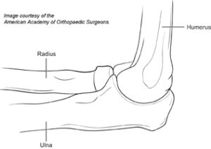
5 name the arteries and nerves that supply elbow joint? The elbow is the visible joint between the upper and lower parts of the arm.it includes prominent landmarks such as the olecranon, the elbow pit, the lateral and medial epicondyles, and the elbow joint.the elbow joint is the synovial hinge joint between the humerus in the upper arm and the radius and ulna in the forearm which allows the forearm and hand to be moved towards and away from the body. Soft tissue dissection of the ulnar soft tissues shows the ulnar collateral ligament (arrowheads) and the closely apposed flexor digitorum superficialis tendon fibers (arrows). Here we will look in detail about the ligaments, the common injuries affecting them, how they are diagnosed and treated. Distally, it attaches to the annular ligament of the radius and coronoid process of the ulna 1,3,5. The latter could be confused with a superficial layer to the ulnar collateral. The radial collateral ligament, on the lateral elbow, connects the radius to the humerus. It is a part of the lateral (radial) collateral ligament complex and located at the posterolateral aspects of the elbow joint. The radial collateral ligament is found on the lateral side of the joint, extending from the lateral epicondyle, and blending with the annular ligament of the radius (a ligament from the proximal radioulnar joint). Being a hinge joint, the only movements allowed by the elbow are flexion and extension of the joint and. Elbow joint and radioulnar joints dr. Superior depiction of muscles, ligaments, and tendons as well as the ability to directly visualize nerves, bone marrow, and hyaline cartilage are advantages of magnetic resonance imaging relative to conventional imaging techniques. Lateral ulnar collateral ligament (lucl):
The articular surfaces are covered with hyaline cartilage. Figure 10.3 ulnar collateral ligament dissection. This capsule surrounds the elbow joint and contains lubricating fluid called synovial fluid. The latter could be confused with a superficial layer to the ulnar collateral. It is am important stabiliser of the proximal radioulnar and radiocapitellar joint.
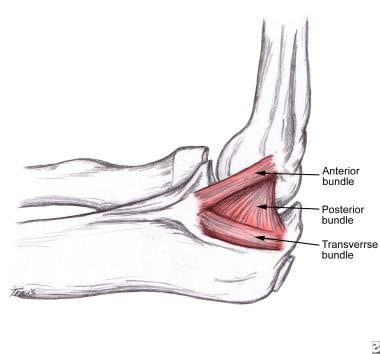
Elbow Joint Anatomy And Significance Bone And Spine from boneandspine.com The ulna collateral ligament, on the medial side of the elbow, connects the ulna to the humerus. It extends from the medial epicondyle of the humerus to ulnar tuberosity on the medial aspect of the coronoid process and the medial aspect of the trochlear notch/olecranon of the ulna. Elbow joint and radioulnar joints dr. Ligaments are bands of tissue that connect one bone to another to form the joints. The humeroulnar and the humeroradial joints each have a ligament connecting the two bones involved at the articulation: A brief description of the ligaments of the elbow joint. 6.1 subluxation of head of radius/pulled elbow; An ulnar collateral ligament (ucl) tear is an injury to a ligament in your elbow.
A brief description of the ligaments of the elbow joint.
These ligaments allow for movement and stretching of the elbow while resisting dislocation of the bones. An elbow sprain is when you stretch or tear one, two or all three ligaments which provide support for your elbow. The annular ligament of the elbow extends from the ulna around the head of the radius to hold the bones of the lower arm together. 1 describe the type and articular surfaces of elbow joint. The radial collateral ligament, on the lateral elbow, connects the radius to the humerus. 3 ligaments of the elbow joint. Ligaments are bands of tissue that connect one bone to another to form the joints. Soft tissue dissection of the ulnar soft tissues shows the ulnar collateral ligament (arrowheads) and the closely apposed flexor digitorum superficialis tendon fibers (arrows). The joint capsule of the elbow surrounds all 3 joints. 3 describe the movements of elbow joint. Ligaments the joint capsule of the elbow is strengthened by ligaments medially and laterally. An ulnar collateral ligament (ucl) tear is an injury to a ligament in your elbow. The ucl attaches the humerus to the ulna, which helps support and stabilizes your arm.
The humeroulnar and the humeroradial joints each have a ligament connecting the two bones involved at the articulation: 3 ligaments of the elbow joint. When the ligament is injured, it could be stretched, partially torn, or completely torn. 2 describe the capsule and ligaments of elbow joint. The ligaments which hold your elbow bones together are the medial, lateral and annular ligaments.
Elbow Treatment Overview Genesis Orthopedics Sports Medicine from genesisortho.com Superior depiction of muscles, ligaments, and tendons as well as the ability to directly visualize nerves, bone marrow, and hyaline cartilage are advantages of magnetic resonance imaging relative to conventional imaging techniques. It is a part of the lateral (radial) collateral ligament complex and located at the posterolateral aspects of the elbow joint. The articular surfaces are covered with hyaline cartilage. The ucl attaches the humerus to the ulna, which helps support and stabilizes your arm. The radial collateral ligament also contributes to posterolateral rotational stability. The radial collateral ligament is found on the lateral side of the joint, extending from the lateral epicondyle, and blending with the annular ligament of the radius (a ligament from the proximal radioulnar joint). When the ligament is injured, it could be stretched, partially torn, or completely torn. A brief description of the ligaments of the elbow joint.
Soft tissue dissection of the ulnar soft tissues shows the ulnar collateral ligament (arrowheads) and the closely apposed flexor digitorum superficialis tendon fibers (arrows).
The last two ligaments connect the radius to the ulna, and are the anular and quadrate ligaments. Occurs between the trochlea and capitulum of the humerus and the trochlear notch of the ulna and the head of the radius. The radial collateral ligament is found on the lateral side of the joint, extending from the lateral epicondyle, and blending with the annular ligament of the radius (a ligament from the proximal radioulnar joint). Lateral epicondyle to annular ligament. 2 describe the capsule and ligaments of elbow joint. Here we will look in detail about the ligaments, the common injuries affecting them, how they are diagnosed and treated. It extends from the medial epicondyle of the humerus to ulnar tuberosity on the medial aspect of the coronoid process and the medial aspect of the trochlear notch/olecranon of the ulna. The major static elbow stabilizers are the medial (ulnar) and lateral (radial) collateral ligaments and the ulnohumeral joint. 1 describe the type and articular surfaces of elbow joint. This capsule surrounds the elbow joint and contains lubricating fluid called synovial fluid. Coronoid fossa fat pad (anterior) radial fossa fat pad (anterior) The annular ligament of the elbow extends from the ulna around the head of the radius to hold the bones of the lower arm together. Ligaments the joint capsule of the elbow is strengthened by ligaments medially and laterally.
An ulnar collateral ligament (ucl) tear is an injury to a ligament in your elbow 3. liga. Being a hinge joint, the only movements allowed by the elbow are flexion and extension of the joint and.

Komentar
Posting Komentar